

Mitochondria: everyone’s favorite organelle. The “powerhouse of the cell” is vital to basically every function your body performs and even has its own genome. For at least a century, scientists had observed that this organelle occasionally does something weird – its membrane reshapes itself to look a bit like beads on a string. No one knew why it was happening and it was dismissed as a bit of an oddity. Until now.
The rest of this article is behind a paywall. Please sign in or subscribe to access the full content.Ancient bacterial leftovers
Mitochondria exist in every cell in your body except red blood cells. They are tiny, energy-generating factories wrapped up in a membrane, and they contain their own unique set of chromosomes, which you typically only inherit from your mother.
Through a series of highly complex biochemical pathways, mitochondria produce adenosine triphosphate (ATP), the body’s energetic currency that cells need to thrive. In cell biology terms, they’re kind of a big deal.
It’s probably surprising, then, that there are still mitochondrial mysteries left to uncover. That’s why we were so intrigued to learn about a new study that is finally answering a question about mitochondria that has existed for over 100 years. We sat down with first author Dr Juan C. Landoni from the Federal School of Technology in Lausanne, Switzerland, to find out more.
“Mitochondria have their own DNA, they have their own genome, which is left over from when they were bacteria,” Dr Landoni told IFLScience.
We should probably talk about that real quick – according to the most widely accepted theory of the origins of complex life, an ancient microbe called an Asgard archaeon once swallowed a bacterium. Rather than being digested, the bacterium thrived and eventually formed what we now know as a mitochondrion. This is all thought to have happened 1.6 to 2.2 billion years ago.
The mitochondria we know and love today have a genome that only encodes about 13 proteins, Dr Landoni explained, “but they're all completely crucial, like fundamental for human health”. They also need to be organized carefully, because these proteins are required throughout the organelle but can’t move around easily.
There’s also the problem of making new mitochondria – “you have to always make a mito from another mito,” Dr Landoni explained, but you can’t have a situation where your new mitochondrion only has half the genes it needs.

When a whole cell divides to produce daughter cells, we understand that process in granular detail. But when mitochondria divide, scientists have very little clue how it happens
Pearls on a string
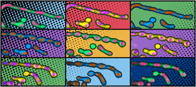
In order to maintain proper organization of the mitochondrial DNA (mtDNA), each cell contains hundreds to thousands of copies packaged into structures called nucleoids, which are regularly spaced along the mitochondrial membrane. Under certain conditions, as you can see in the animation above, the membrane squishes itself into a new conformation that looks like beads on a string.
This is called “pearling”. Dr Landoni told us he has spoken to many scientists who have seen it happen under a microscope but have assumed it was a weird fluke, or a sign that the mitochondria weren’t happy.
“[I call mitochondria] the blessing and the curse because they’re extremely sensitive to anything,” he said. “So if I see healthy mitos, that means the cell is very happy. And I know that I'm not working with a stressed cell because the mitos immediately, as soon as anything happens, they will fragment and they will start behaving weirdly.”
In the course of their research, he and the team were even able to go back to some of the earliest observations of pearling, buried in lab notebooks from the turn of the 20th century belonging to pioneering cell biologist Margaret Reed Lewis. She and her husband published many detailed observations and drawings of a range of cellular functions.
All these years later, it took a team from across scientific disciplines bringing together their perspectives to finally figure out just how the mitochondria achieve the pearling that the Lewises saw through their microscope.
Physics is the key
The breakthrough came when some of Dr Landoni’s physicist colleagues insisted that what they were seeing in the mitochondria wasn’t a biologically driven process, but a physical one. Pearling, they declared, would happen by itself, with or without any proteins present – because physics.
“It was quite hard to convince biologists that, oh no, there is no protein. Like there are proteins regulating this, there's proteins triggering it or enabling it to happen. But the actual fact of the pearling happens spontaneously and likely precedes the mitochondria itself,” Dr Landoni told IFLScience.
That’s a hard pill to swallow for biologists who are used to the idea that cellular processes happen because some upstream factor, like a gene or protein, makes them happen: “How do we study something that just happens because it happens?”
Despite their skepticism, however, the team worked together and eventually was able to reach a fuller understanding of the process than had ever been achieved before.
Pearling is a natural, physical consequence of how mitochondria are structured. Dr Landoni’s biophysicist colleagues investigated this by doing everything from “blasting the cells with a laser, poking them with a stick”, and discovered that “anything can make it happen”. There are, he said, “a million ways” to get the membrane to adopt this shape.
I think the mito kind of adapted its gene expression system to match this regularity.
Juan C. Landoni
But that doesn’t mean the mitochondria are passively letting it happen. Rather, it appears they adapted around this natural process to take advantage of it and evolved various biological mechanisms by which they can wrest back some control over pearling.
“I always get asked, why did this evolve? And it didn't. It was already there,” Dr Landoni explained. “So, I think the mito kind of adapted its gene expression system to match this regularity. Because if you're going to be constantly turned into pearls with this regular distancing, you might as well optimize your region of influence or your gene expression system to have that reach.”
What’s next?
Mitochondria are vital cellular workhorses, but things can and do go wrong. According to the United Mitochondrial Disease Foundation, a child who will go on to develop mitochondrial disease by age 10 is born every 30 minutes.
“Most mitochondrial diseases don't have a clear pathogenic explanation,” Dr Landoni told IFLScience. “The biggest unexplained feature of them is that similar mutations often lead to [symptoms in] completely different constellations of organs.”
Let’s see if the muscle and the liver and the heart use different strategies to maintain their mitochondrial health and mitochondrial DNA distribution.
Juan C. Landoni
You might expect that if one mtDNA mutation in a patient affects the heart, that same mutation would also affect the heart in someone else, but that isn’t the case – “and we have no idea why”. Perhaps it’s something to do with pearling. Dr Landoni said one hypothesis is that different organs could use slightly different genome organization strategies, so the more we understand about this process, the better equipped we will be to research these diseases.
Part of the reason why pearling wasn’t properly understood before was that we didn’t have the technology to observe it in a high enough level of detail. Now that technology has moved on, Dr Landoni is keen to pursue imaging in whole organisms, rather than just cell cultures, to observe pearling as it happens.
“Let’s see if the muscle and the liver and the heart use different strategies to maintain their mitochondrial health and mitochondrial DNA distribution. And how does that directly impact health and mitochondrial diseases specifically?”
“I need to change projects because this looks cool”
This story reads like a throwback to “old-school” scientific discoveries: scientists saw something they couldn’t explain and were inspired to keep digging until they figured out what was going on.
“I think the most exciting part was when I first saw it, because – it feels fake – but I literally went to the office of my boss and was like, I need to change projects because this looks cool, and she trusted me on it,” Dr Landoni told IFLScience.

“There was like this kind of joining of different people that have observed it but didn't know what to do with it. And then, together, we did something with it. So that felt good as well. It felt [like] very traditional science from back in the day of sharing these small pieces of information and trying to gather something.”
And who knows what other mysteries could be waiting in notebooks like Lewis’s, just waiting for modern technology to catch up and for someone to have the idea of going back to them?
“It took 100 years for one of those drawings to actually be paid enough attention to find a function, and who knows how many of the other wiggles that she made actually will eventually turn into something,” Dr Landoni said. “So that was just really neat.”
The study is published in Science.